The AO Spine North America Research Committee (AO SNARC) congratulates the recipients of the 2025 Young Investigator Research Grant Award (YIRGA).
The AO SNARC continues to offer research funding that is available to new investigators (within 5 years of first appointment). The purpose of these grants is to encourage high-quality, clinically-relevant spinal or spinal cord research by providing start-up funding of up to $20,000 for one year.
About This Year’s Early Career Spine Research Funding Winners
This year’s award recipients are exploring a range of topics including the potential application of AI to improve early detection and surveillance of cervical myelopathy; identifying risk factors and ways to reduce poor cognitive outcomes for elderly patients undergoing spine surgery; addressing the high rate of pedicle screw failure in osteoporotic patients through a combination of a flexible pedicle screw instrumentation with robotic-guided curved drilling trajectories; and harnessing digital technologies such as smartphones and wearables to assess and monitor degenerative cervical myelopathy.
Learn more about these surgeons and their work below.
Mark your calendars! The 2026 YIRGA application period opens this fall. View application requirements here.
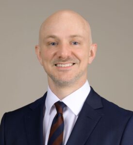 Dr. Joseph Drain
Project Title: Automated Detection of Cervical Spine Canal Diameter and Anatomic Structure
Instagram | LinkedIn
Dr. Joseph Drain, assistant professor and spine surgeon at The University of Texas, Houston, has been awarded one of the 2025 AO Spine North America Young Investigator Research Grants. His research project, titled “Automated Detection of Cervical Spine Canal Diameter and Anatomic Structure,” aims to lay the foundation for a long-term research career in medical imaging and AI.
“This isn’t just a wave, it’s a change in sea level,” Dr. Drain says of the role artificial intelligence will play in the future of medicine. His current project uses machine vision to calculate standard cervical canal diameter values from normal CT scans. By leveraging machine learning, he can analyze not just tens or hundreds, but thousands of scans in a consistent, automated way.
Dr. Drain’s goal is to improve early detection and surveillance of cervical myelopathy. CT scans are widely available, quick to obtain, and frequently ordered in trauma settings, often without specific suspicion of cervical pathology. “We now have the tools to estimate population norms based on large-scale datasets,” he explains.
He hopes this project will be the first of many, building a robust pipeline of collaborators, techniques, and expertise focused on automated image processing and pathology recognition.
Dr. Drain credits AO North America for providing essential support: “Their backing is the gasoline that powers the engine,” he says. He’s committed to bringing AI into orthopaedics and getting more orthopaedic surgeons involved directly in data-driven innovation.
 Dr. Mark Lambrechts
Project Title: Short- and Long-Term Cognitive Effects Fo lowing Anesthesia for Spinal Surgery: A Prospective Observational Study
Dr. Mark Lambrechts, assistant professor and spine surgeon at Washington University in St. Louis, has been awarded a 2025 Young Investigator Research Grant Award for his study, “Short- and Long-Term Cognitive Effects Following Anesthesia for Spinal Surgery: A Prospective Observational Study.”
Dr. Lambrechts’s research focuses on how spinal surgery impacts cognitive function, particularly in elderly patients. While smaller procedures are generally well tolerated, longer surgeries carry a higher risk of cognitive decline, including short-term memory loss and potential links to dementia.
“What’s unclear is how a patient’s individual biology, especially their proteomics, affects their ability to tolerate surgery,” Dr. Lambrechts explains. The goal is to better understand these risks and provide clearer guidance for patients.
As the aging population continues to grow, more older adults are undergoing spine surgery. Identifying risk factors for poor cognitive outcomes is becoming increasingly critical. This study aims to uncover those risk factors and explore ways to reduce complications.
“A unique aspect of our research is analyzing patients’ protein markers, which have been linked to dementia in the general population,” says Dr. Lambrechts. “This may help us identify patients at high risk for cognitive decline before surgery, allowing for better planning and possible interventions.”
Ultimately, the long-term goal of the project is to pinpoint preoperative markers that can predict cognitive decline, and eventually develop therapies to address them. If certain proteins are found to contribute to this risk, targeted treatments may be possible in the future.
Dr. Lambrechts emphasizes the importance of support from AO North America: “Without their funding, this study wouldn’t be possible due to the high cost of proteomic analysis. Their support is vital to advancing this research.”
 Dr. Jordan Amadio
Project Title: Morphable Pedicle Screw Technology for Reducing Complications in Minimally Invasive Spinal Fixation
Dr. Jordan Amadio is Clinical Affiliate Faculty at Dell Medical School and Affiliated Faculty at UT Texas Robotics, University of Texas at Austin. He is a practicing neurosurgeon and serves as Chief of Spinal Neurosurgery at Olympia Neurological Institute.
Dr. Amadio is leading an innovative project focused on the development of Spatial Spinal Fixation, a groundbreaking approach that combines a novel design for flexible pedicle screw instrumentation with robotic-guided curved drilling trajectories. This work addresses a critical challenge in spinal surgery: the high rate of pedicle screw failure in osteoporotic patients, where traditional rigid screws can loosen in up to 50% of cases.
His team at UT Austin is engineering flexible pedicle screws designed to morph within the vertebral anatomy, distributing mechanical loads along continuous surfaces rather than relying on discrete contact points. This technology is paired with a Concentric Tube Steerable Drilling Robot, capable of navigating curved, J-shaped trajectories to reliably target regions of high bone mineral density, something not achievable with existing straight-line drilling techniques.
With over 500,000 spinal procedures performed annually in the U.S.—at a cost of $13 to $40 billion and osteoporotic fractures occurring every 20 seconds, spinal care is facing a growing crisis. The current generation of pedicle screw technology, largely unchanged since the 1990s, is being pushed to its limits by increasingly complex patient populations with compromised bone quality.
This research aims to overcome four fundamental limitations in current spinal instrumentation: anatomical constraints, rigid drilling tools, poor bone targeting, and stress concentration. By enabling surgeons to place flexible implants in optimal bone regions through curved trajectories, Dr. Amadio’s work could significantly reduce the rate of revision surgeries and improve outcomes for millions of patients who are currently underserved.
Dr. Amadio’s motivation stems from his daily experience in the operating room, where the limitations of current spinal fixation technology are most evident, especially in high-risk populations such as osteoporotic or cancer patients. “Our ‘surgineering’ collaboration with Dr. Alambeigi at UT Texas Robotics began when we recognized that the spine’s complex anatomy demands non-rigid implants that exploit natural geometry,” he explains. “We realized the fundamental problem isn’t just screw design we’ve been constrained to straight-line thinking in a curved anatomical world. That insight led us to envision spatial fixation as a paradigm shift.”
Within the next few years, the team’s technology could enable patient-specific biomechanical planning using quantitative CT (QCT) scans, followed by execution of curved drilling paths that reach optimal bone regions with sub-millimeter precision. Long-term, this could establish a new surgical standard in which flexible implants and robotic guidance become the norm for treating complex spinal conditions. The platform also holds promise for applications in bone tumor surgery and orthopedic joint reconstruction.
The project’s next phase is being catalyzed by the AO Spine Young Investigator Research Grant, which supports a focused 12-month effort to achieve comprehensive biomechanical validation of flexible pedicle screws using benchtop and cadaveric models. This critical data will enable clinical translation
. The foundation’s support also provides essential validation for attracting federal funding and building industry partnerships. The grant positions the team to scale their transformative technology from the lab to real-world patient care.

Dr. Jetan Badhiwala
Project Title: The use of digital health technology for a personalized medicine approach to degenerative cervical myelopathy
Dr. Jetan Badhiwala, assistant professor and spine surgeon at The University of Toronto has been awarded one of the 2025 AO Spine North America Young Investigator Research Grants. This project investigates the potential of digital health technologies to transform the assessment of adult patients with degenerative cervical myelopathy (DCM). The aim is to determine whether these tools can help define personalized disease profiles that predict quality of life and functional outcomes, while also identifying new outcome measures that are sensitive to changes in the disease over time.
Digital technologies such as smartphones and wearable devices are now ubiquitous, intricately woven into the fabric of modern life. These tools continuously and passively capture vast amounts of health-related data through everyday physical, social, and behavioral activity. They also serve as platforms for actively collecting health-related measures. Wearable sensors, for example, can track physiological signals, movement, gait, and sleep patterns without requiring user input. Meanwhile, smartphone usage generates data on gross motor activity, communication dynamics, social interactions, speech, and cognitive function, rich information that can be captured using passive digital phenotyping.
Smartphones also support active methods of data collection, such as ecological momentary assessments (EMAs) brief, repeated surveys that gather real-time information about symptoms and subjective experiences in the user’s natural environment. Together, these tools offer a unique opportunity to collect objective and subjective health data in real-world settings and in real time.
The growing application of digital health technologies across medical fields points to their potential in improving both patient evaluation and care. In the context of DCM, their integration could be especially powerful. These technologies make it possible to obtain reliable, longitudinal, and multidimensional measures of function, both objective and subjective, on a large scale. Such data may help characterize different disease subtypes, reveal biomarkers of disease severity, and offer prognostic insights into how a patient might respond to treatment. They could also serve as entirely new outcome measures that are more accurate and reflective of the patient’s lived experience.
Current tools used to assess DCM, such as the modified Japanese Orthopaedic Association (mJOA) scale, present several limitations. These measures are subjective, depending heavily on patient self-reporting, and narrowly focused, assessing only select areas of functional impairment. Furthermore, they are usually conducted during clinic or research visits, which are cross-sectional and prone to recall bias. These methods fail to account for how symptoms and function vary over time or across different real-life contexts.
The few existing objective tools, like the GRASSP-M, are task-based and limited to one-time evaluations conducted in controlled settings. This means they also miss the day-to-day dynamics of DCM and the complex interplay of symptoms as they fluctuate. There remains a critical unmet need for assessment tools that offer continuous, real-time, objective and subjective insights across a broad range of functional domains in real-world environments.
This project’s goal is to develop a novel, digital approach to assessing and monitoring DCM through the use of smartphones and wearables. By capturing multidimensional data over time, the research seeks to generate clinically meaningful information that can support decision-making and ultimately become part of standard clinical practice.
Support from AO North America will be instrumental in launching this pilot study. This initial effort will inform a larger grant application and a future prospective study, while also increasing visibility and opportunities for knowledge translation. Through this work, the hope is to bring cutting-edge digital innovation to patients with DCM, deepening understanding of the disease and enhancing care at every stage.

